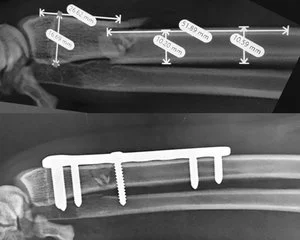
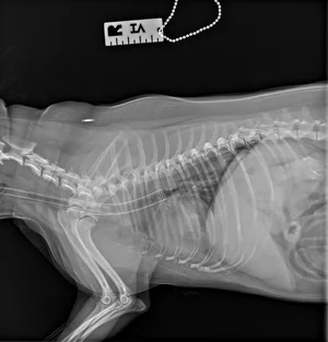
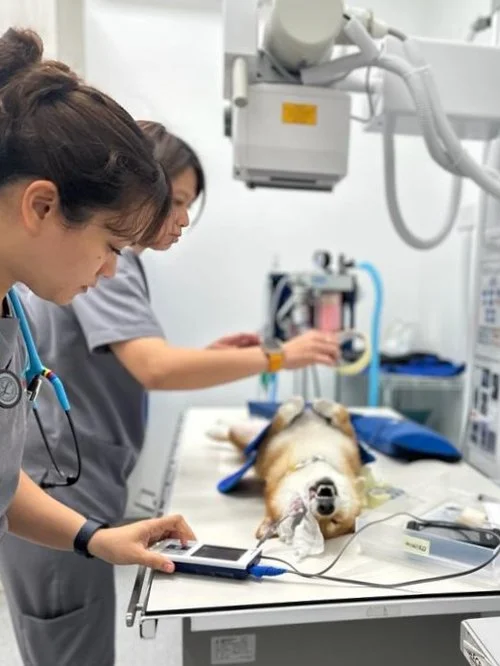


Diagnostic imaging services are essential for animals to have accurate and early diagnosis of medical conditions in various fields, including cancer, orthopaedics, heart health, neurology, and surgical evaluations. They play a pivotal role in facilitating treatment planning and allowing our specialists, veterinarians, and surgeons to precisely understand the extent and location of issues. Diagnostic imaging services also support preventive care by detecting hidden issues before they become symptomatic.
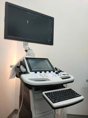
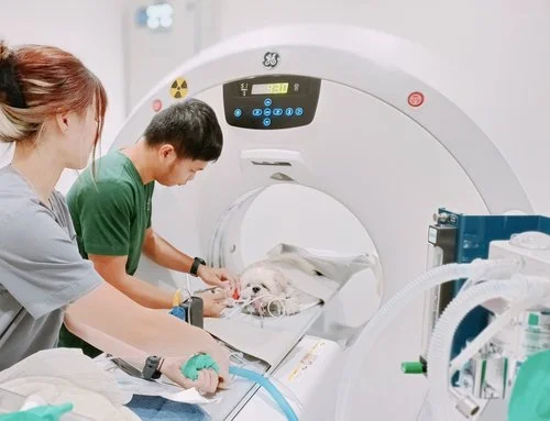
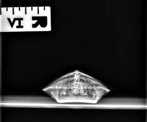
The Diagnostic Imaging and Radiology Service at the Beecroft uses advanced imaging technologies such as radiography (x-ray), ultrasound and computed tomography (CT) for the diagnosis, prognosis, and treatment planning of diseases in companion and exotic animals. Our 16 slice detector creates 32 slice using specialized software to give high-resolution images.
Diagnostic imaging is completely non-invasive and it allows thorough examination of specific areas of an animal’s body, especially internal organs. The high-clarity images allow us to see the precise problem and to make a precise diagnosis.
Unparalleled Diagnostic Precision
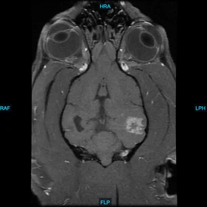
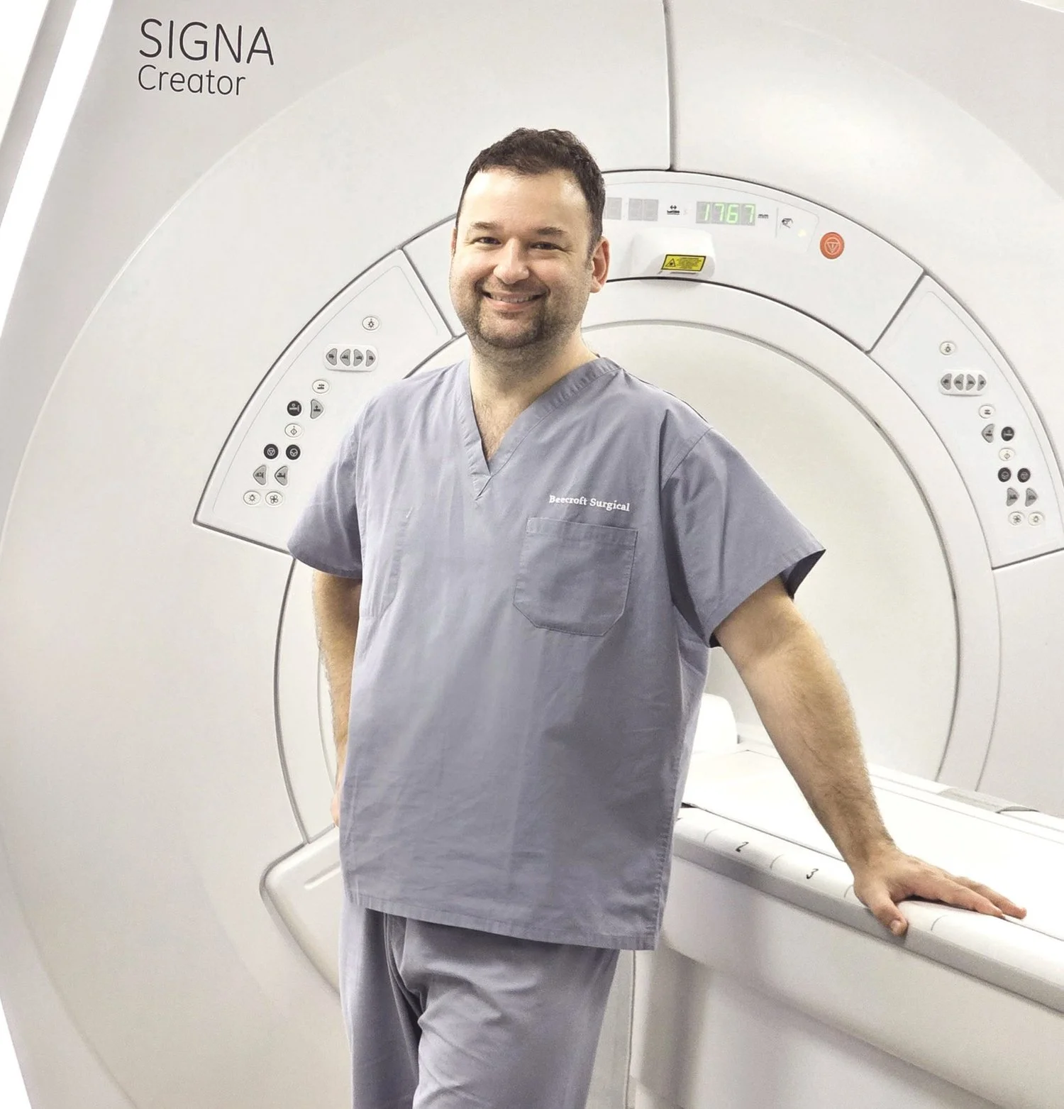
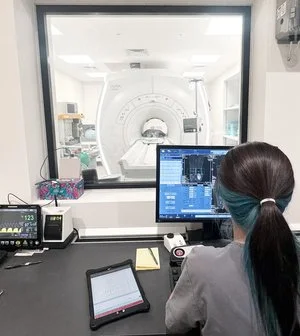
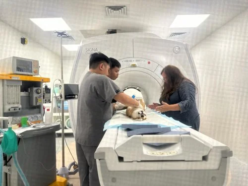
Our new high-field MRI (GE Signa, 1.5T) is a game-changer for diagnosing complex neurological and orthopaedic conditions of pets. Unlike traditional CT scans which excel in certain scenarios, MRI offers superior soft tissue and multi-planar imaging capabilities, allowing us to visualise intricate structures with unmatched clarity.
Precision in Neurological Diagnoses
For cases involving neurological conditions such as seizures, encephalopathies, or spinal cord diseases, our high-field MRI provides detailed insights into the central nervous system. This enables us to pinpoint the exact location and nature of abnormalities, facilitating precise treatment planning.
Orthopaedic Excellence
In orthopaedic cases such as ligament injuries, joint diseases, or unexplained lameness, our high-field MRI delivers exceptional diagnostic accuracy. It helps identify soft tissue injuries, articular cartilage disorders, and musculoskeletal abnormalities that might go undetected with other imaging modalities.
Here are some conditions where High-Field MRI shines (and where a CT-scan would less likely identify a lesion):
Diagnosis of epilepsy:
Our hospital has a wide range of diagnostic equipment available to help with diagnosis:
Minimally invasive techniques are currently being investigated in veterinary medicine and have the potential to provide alternatives for our patients in whom conventional therapies are declined, not indicated, or associated with excessive morbidity or mortality.
Interventional radiology can be applied to any body system, in patients of all sizes, and therefore will likely have very broad applications in our patients.
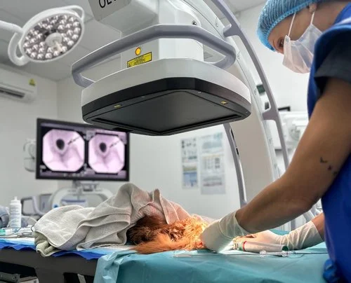
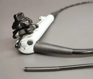
Endoscopy, in combination with fluoroscopy, can be used for various minimally invasive interventions as well. This modality is termed interventional endoscopy (IE). Some examples include endoscopic ureteral stenting, percutaneous nephrolithotomy (PCNL), cystoscopic-laser ablation (CLA) of ureteral ectopia, aberrant turbinates ablation for BOAS, urethral Transitional Cell Carcinoma (TCC) ablation and laser ablation myringotomy. These endoscopic procedures are facilitated by the use of fluoroscopy to aid in the guidance of an assortment of guidewires, catheters and stents.